- Review
- Open access
- Published:
Early prediction of Alzheimer's disease using convolutional neural network: a review
The Egyptian Journal of Neurology, Psychiatry and Neurosurgery volume 58, Article number: 130 (2022)
Abstract
In this paper, a comprehensive review on Alzheimer's disease (AD) is carried out, and an exploration of the two machine learning (ML) methods that help to identify the disease in its initial stages. Alzheimer's disease is a neurocognitive disorder occurring in people in their early onset. This disease causes the person to suffer from memory loss, unusual behavior, and language problems. Early detection is essential for developing more advanced treatments for AD. Machine learning (ML), a subfield of Artificial Intelligence (AI), uses various probabilistic and optimization techniques to help computers learn from huge and complicated data sets. To diagnose AD in its early stages, researchers generally use machine learning. The survey provides a broad overview of current research in this field and analyses the classification methods used by researchers working with ADNI data sets. It discusses essential research topics such as the data sets used, the evaluation measures employed, and the machine learning methods used. Our presentation suggests a model that helps better understand current work and highlights the challenges and opportunities for innovative and useful research. The study shows which machine learning method holds best for the ADNI data set. Therefore, the focus is given to two methods: the 18-layer convolutional network and the 3D convolutional network. Hence, CNNs with multi-layered fetch more accurate results as compared to 3D CNN. The work also contributes to the use of the ADNI data set, where the classification of training and testing samples is divided with such a number that brings the highest accuracy achieved with 18-layer CNN. The work concentrates on the early prediction of Alzheimer's disease with machine learning methods. Thus, the accuracy achieved is 98% for 18-layer CNN.
Introduction
The deterioration of physical and neurological functions in persons is part of the aging process. Although deterioration is natural, it can significantly influence some persons due to certain risk factors. Alzheimer's disease is a neurocognitive disorder occurring in people in their middle or old age, and it affects 46.8 million people globally and can impact a person's quality of life [1]. AD populations are estimated to increase to 106.8 million by 2050 [2, 3]. The estimated cost of long-term health care for dementia patients is about $290 billion [4]. Research toward early AD diagnosis is ongoing to slow down the abnormal degradation of neurons in the brain. It also produces emotional and financial benefits for the patient family [5]. This disease causes the person to suffer from memory loss, unusual behavior, and language problems. It is caused due to the tangled bundles of neurofibrillary fibres of the brain and certain regions of the brain like the entorhinal cortex and hippocampus [6]. The initial symptoms, such as episodic memory impairment and the navigational problem of the patient, are typical variants. The higher order symptoms include memory loss, impaired judgment, difficulty in identifying objects, confusion in paying bills and driving a vehicle, and placing objects in odd places.
Alzheimer's disease is divided into three periods: the primary period, the intermediate period, and the last period of dementia. AD is diagnosed through the brain monitoring modalities, such as CT (Computer Tomography) scan and PET (Positron Emission Tomography) scan resting-state functional magnetic resonance imaging (RS-FMRI) [7].
AD is a neurodegenerative disease with symptoms, such as motor dysfunctions of the body. The results of this work created a strong link between inflammation and neurotoxic kynurenines of human samples. A need for biomarkers is necessary due to chronic low-grade inflammation [8].
The dissection of neuroprotective and neurodegenerative components of AD-affected areas in the brain. It focuses on etiology, pathomechanism, biomarkers, Imaging techniques, and novel therapeutic targets of Alzheimer's disease [9].
The EEG biomarkers help predict clinical outcomes in patients regularly. The accuracy of psychophysiological biomarkers based on EEG while predicting the outcome of the patients. The machine learning technique reached an accuracy of 83.3%, with EEG-based functional connectivity predicting clinical outcomes in nontraumatic patients [10].
The neurovisceral integration model of fear is being used. That is, A richer understanding of neurovisceral concomitants of this function has both theoretical and clinical implications [11].
Treatment of fear-related disorders occurs due to neuro disorder AD. To fix this, research has been done using a novel frequency domain analysis of heart rate using a short-time Fourier transform from a point process modeling algorithm [12].
Pavlovian and Instrumental learning can be integrated to guide behavior in a phenomenon experimentally known as Pavlovian-to-Instrumental Transfer (PIT) to investigate numerical applications in clinical contexts such as working memory affected due to AD [13].
Mitochondrial DNA is identified as an inheritable metabolic disease with neurological manifestation and pathogenesis of illness, including neurodegenerative diseases, such as Alzheimer's disease [14].
Kynurenic acid (KYNA) is an endogenous tryptophan (Trp) metabolite with neuroprotective properties. KYNA plays critical roles in nociception, neurodegeneration, and neuroinflammation. A lower level of KYNA is observed in patients with neurodegenerative diseases, such as Alzheimer's [15].
Machine learning
Understanding machine learning and the standard machine learning approaches used in AD prognosis is necessary before starting the deeper examination of machine learning methodologies. Artificial intelligence includes machine learning, which contains various tools for making probabilistic and statistical judgments based on prior knowledge. Classifying new events and forecasting new patterns depends on prior learning (training). When compared to standard statistical methods, machine learning is much more powerful. For machine learning to be successful, it is essential to have a good understanding of the problem and the algorithms' constraints. As a result, it has a fair chance of success if experimentation is carried out appropriately, training is used effectively, and outcomes are rigorously validated.
Background
This paper reviews the state-of-the-art techniques and data sets used to detect Alzheimer's disease. The various researchers' work, different classifiers used to detect Alzheimer's disease early, and the results obtained are discussed. A literature survey is conducted to know every possibility is explored to detect the initial stages of AD using the ML approach. This survey includes a list of methodologies, data sets, and accuracy gained. The study exhibits the most appropriate strategy for quick treatment of AD based on studies conducted from 2016 to 2021.
Mehmood et al. [1] stated that identifying Alzheimer's on magnetic resonance images in the initial period is carried out using mild cognitive impairment detection using the tissue segmentation of the brain with the help of multiple layers called structured deep learning. The study uses Visual Geometry Group architecture belonging to deep convolutional neural network architecture. The FMRI images used in this paper are gathered from ADNI (Alzheimer's disease neuroimaging initiative) and are found online at adni.loni.usc.edu. To analyze and verify the development of MCI, i.e., mild cognitive impairment, different biomarkers such as structural MRI, PET, and MRI were analyzed and authenticated to detect the traces of AD. 300 MRI subjects were considered and further classified as Alzheimer's, late mild cognitive, and initial mild cognitive periods. The techniques used in this study are CNN with a multi-layered form of various layers, such as convolution layer, pooling layer, and softmax layer. An accuracy of 98.73% is achieved using multi-layered CNN without data augmentation.
Odusami et al. [16] proposes a deep learning method to detect the early stage of AD. He has proposed a modified ResNet18 model for extracting the features of neuroimaging data from structural magnetic resonance imaging. The data set is fetched from ADNI accessed on January 2021. The data sets are available in DICOMM file format. A total of 413 subjects were considered for the study. The six categories of the database are normal healthy period, light cognitive inability EMCI and notable remembrance, and Alzheimer's is addressed. The techniques used are residual network with 18 layers CNN is proposed. It uses a 3 × 3 seiver, and the phase 1 pooling layer has a 1 × 1 seiver, a completely interlinked, and a softmax layer. The fine-tuned CNN of 18 layered neural networks obtained a separation rate of accuracy of about 99.09%. CNNs are used to detect active magnetic resonance imaging scanned sheets of Alzheimer affected persons. The process is carried out in data collection, preprocessing and fine-tuning, and classification and evaluation stages.
Venugopalan et al. [17] proposed removing noise from MRI scans using automatic encoders for evoking properties from given data. He has stated a novel method of 3D CNN for imaging data, and he concentrated on the hippocampus brain area and features. Audio oral tests are extracted. The ADNI data set is used considering biological markers MRI, PET, and neuropsychological assessments to measure the progression of mild cognitive impairment. The cross-section Magnetic Resonance scan image gathered about 8209 voxels scattered in 18 parts. A total of 220 patients were considered for the test. The total count of MRI images is 503 in number, SNP is 808 in number, and HER is 2004. A three-tier automatic encoder is used with 199,99, and 51 nodes separately for every part. The first step is to filter noise, and the next is to extract 1680 common features and convert input data into 0's and 1's format by shot encoding. An accuracy of 78% is achieved.
Pradhan et al. [18] proposed the detection of different stages of AD. The method used is VGG19 and DenseNet169 architecture for classification. The data set is taken from an open online data set library called Kaggle. There are 6000 images labeled as mild, moderate, very mild, and non-demented AD. The features are considered for 80% of learning and 20% of examining phases. VGG19 has around 10–16 convolutional neural network layers. For image classification, DenseNet is used. Here, VGG19 performs better than DenseNet accuracy of 94% is achieved.
Shah et al. [19] hard and soft voting algorithms were implemented to classify and identify the initial AD period. The data set consists of 437 patients aged between 60 and 96. Among these, 72 people are non-demented, 64 are demented, 70% are used to train the algorithm, and 30% are used to test the algorithm. The classification algorithms are hard voting, soft voting classifiers, and decision tree. SVM is used as a classification method. An accuracy of 84% is obtained for the voting classifier algorithm.
Huanhuan et al. [20] proposed detecting early stages of dementia (ConvNets) with the help of MRI. The classification of scan images is done using gray color regions and white color regions in the scanned images of the brain. The data are collected from the ADNI database. The number of MRI images collected is 615 in number. The data are segregated in the proportion of 3:1:1. Statistical parameter mapping is used in the preprocessing stage to reduce the patient's head movement, and images are reduced to size 192 × 192 × 160. The techniques used for the detection are based on classifiers eResidual Network of 50 layers, eNeural Architecture Search Network. Adding a dropout layer addresses the overfitting problem to the fully interconnected layer. The accuracy rates are separately obtained from around 97.65% to 88.37% for MCI AD.
Razavi et al. [21] highlighted using unsupervised feature learning, which has two steps. The first step is to extract features from the raw data. The methods used are scattered filtering and uncontrolled neural layer network. Sparse filtering and regression are called softmax to classify healthy and unhealthy persons. A few unsupervised learning techniques, such as Boltzman machines and dispersed coding, are used to distribute collected data. The data set used in this method is ADNI with cerebrospinal fluids. The total number of AD patients is 51, and 43 patients have mild traces of suffering from AD. The MRI data were obtained using 1.5T scanners. The highest accuracy obtained is 98.3% while using the softmax regression.
Islam et al. [22] work on AD uses deep learning CNN to analyze brain MRI images. This work also identifies the different stages of the disease. The method also works well with the imbalanced data set. The CNN uses four layers: deep neural layers, batch processing layer, pooling layer, and ReLU layer. According to the 3D brain, MRI data architecture, inception -v4, and Resnet classify the data. The data set used in OASIS has 416 data samples. The training and testing data set is divided into the 4:1 proportion. The performance rates of inception -v4 and Resnet precision rates are 0.81 and 0.82, respectively.
Islam et al. [23] stated that 3D convolutional neural networks work better in visualizing medical images. Brain PET scans are used to detect Alzheimer's disease using 3D CNN, and five visualization techniques are applied. The data set is collected from ADNI (adni.loni.usc.edu). A total of 1230 PET scans of AD patients are available. The applied visualization techniques are guided by Backpropagation Brain area Occlusion and layerwise relevance propagation. 80% of the data set is used for training, 20% for testing, and the remaining 10% for validation. The visualization techniques are used to enhance and focus on the regions of the brain, such as the frontal mid, precuneus, postcentral, temporal mid, and precentral areas. Hence, the system achieved an efficient classification accuracy of 88.76% is achieved.
Thakare et al. [24] stated using EEG to detect Alzheimer's disease. The EEG database is extracted from Kashi Bhai Hospital, Pune, and nineteen numbered channels of the EEG database. First, the patients are diagnosed with a clinical diagnosis of MSME. Based on this, the patients are divided into healthy and AD patients. The EEG signals obtained are converted into a.mat file, and the acquisition is made using Simulink. The features extracted from these EEG waves are mean, standard deviation, and mode using wavelet transforms. The classification uses a support vector machine and a normalized minimum distance (NMD) classifier algorithm. An accuracy of 95% is achieved using SVM holds good as compared to the NMD classifier.
Noor et al. [25] the most popular DL techniques have been explored in detecting those three leading neurological disorders from the MRI scan data. DL methods for the classification of neurological disorders found in the literature have been outlined. The pros, cons, and performance of these DL techniques for the neuroimaging data have been summarized. Prime observation of this study included the maximum usage of CNN in the detection of Alzheimer's disease and Parkinson's disease. On the other hand, DNN has been used with greater prevalence for schizophrenia detection.
Su et al. [26] Magnetoencephalography (MEG) has been combined with machine learning techniques to recognize Alzheimer's disease (AD), one of the most common forms of dementia. A bimodal recognition system based on an improved score-level fusion approach is proposed to reinforce the interpretation of the brain activity captured by magnetometers and gradiometers. This preliminary study found that the markers derived from the gradiometer tend to outperform the magnetometer-based markers. Interestingly, out of the ten regions of interest, the left-frontal lobe demonstrates about 8% higher mean recognition rate than the second-best performing region (left temporal lobe) for AD/MCI/HC classification.
In clinical practice, several standardized neuropsychological tests have been designed to assess and monitor the neurocognitive status of patients with neurodegenerative diseases, such as Alzheimer's disease. Have presented a robust framework to (i) perform a threefold classification between healthy control subjects, individuals with cognitive impairment, and subjects with dementia using different cognitive indexes and (ii) analyze the variability of the explainability SHAP values associated with the decisions taken by the predictive models [27].
This study aimed to determine the influence of implementing different ML classifiers in MRI and analyze the use of support vector machines with various multimodal scans for classifying patients with AD/MCI and healthy controls. Conclusions have been drawn in terms of employing different classifier techniques and presenting the optimal multimodal paradigm for AD classification [28].
Analyzing magnetic resonance imaging (MRI) is a common practice for Alzheimer's disease diagnosis in clinical research. Detection of Alzheimer's disease is exacting due to the similarity in Alzheimer's disease MRI data and standard healthy MRI data of older people. The proposed network can be very beneficial for early stage AD diagnosis. Though the proposed model has been tested only on the AD data set, we believe it can be used successfully for other classification problems in the medical domain [29, 30].
Methods
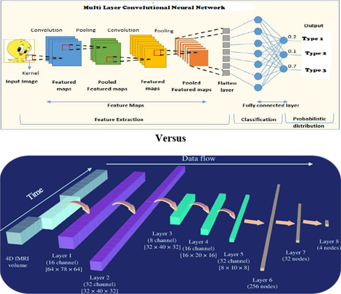
A convolutional neural network with multi-layers such as pooling, softmax regression, and completely interconnected layers is used to detect the disease. A CNN increases the size of the images in length and breadth while decreasing the complexity of the image. A pooling process reduces the overfitting problem as the amount of computation and parameters are reduced. The transfer learning model with customized VGG architecture is used to get the highest accuracy rates [1]: the data collection, preprocessing, fine-tuning, and classification stages. Fine-tuning is used to reduce errors with the help of ImageNet. It uses the residual network with optimal parameters ReLU and stochastic gradient descent. A novel deep learning method with shallow models for integrating data and autoencoders in a minimal data set. VGG 19 of 16 convolutional layers when a large data set is available to classify, and the dense net is utilized to reduce the number of parameters [17].
DenseNet 169 is used for image classification. Both models are compared; VGG 19 performs better than DenseNet [18]. Used the support vector machines for classification and used hard and soft voting classifiers to get the optimum accuracy and use of decision trees for regression, making the system fast and efficient in predicting the missing values of the field [19]. ConvNet for classification and ensemble machine learning techniques are used for a final product from CNN layers. Backpropagation networks either increase or decrease weights to match output with input. Bernoulli's function is used to avoid the overfitting problem [20]. Sparse filtering and softmax regression are trained automatically to identify healthy and unhealthy individuals. These two methods are called the two-stage learning method [21]. These are used for deep convolutional neural networks with four functions, pooling, convolution, batch normalization, and rectified linear unit [22]. The techniques such as 3D convolutional neural networks and visualization techniques include layerwise relevance propagation, guided backpropagation, and sensitivity analysis to detect AD [23]. Proposed the use of support vector machines and normalized minimum distance classifier, and it uses a supervised learning model for better results [24]. Table 1 illustrates the list of methodologies.
Comparison of 18-layer CNN and 3D CNN
The two main components of the CNN architecture are a toolkit that analyses and identifies the properties of the image with a process called feature extraction and a second component based on the prediction process, which estimates the image category from the previous stages. A total of five layers are used: CNN layer, max-pooling layer, completely inter-connected layer, activation layer, and dropout layer. This set of five layers is expanded with approximately 240 filters, each of size 5 × 5. The input for these CNN layers is FMRI image, Pet, and CT scan images that undergo all the preprocessing and conversion processes in the proposed methodologies. In the case of 18-layered CNN, the model predicts the output with the highest accuracy as it has to pass through all the bitwise filters. Hence, with 3D CNN networks, the detection of AD disease might be restricted to less accuracy than 26-layered CNN. Detecting damaged neurofibrils in the brain is easily verified with the multi-layered CNN. In the survey of these related works, maximum use of CNN is being done. Figure 1 represents the comparison of multi-layered CNN versus 3D CNN.
Data set
The data set used in the following papers is shown in Table 2, along with the description data set source, the total number of samples used, the count of training and testing samples, and the number of NC—Normal Control, LMCI—Late Mild Cognitive Impairment, EMCI—Early Mild Cognitive Impairment, and Alzheimer's disease.
Results
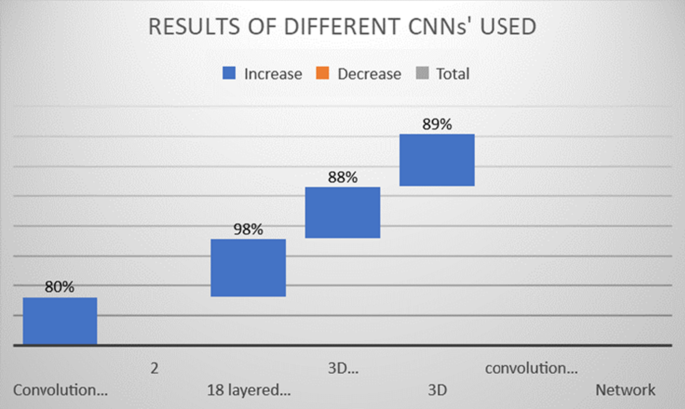
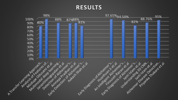
This section illustrates the results and outcomes of various works shown in Tables 3, 4, and 5 and Figs. 2 and 3.
According to the different works examined, the detection of AD carried out using ResNET18 networks holds the highest accuracy of 98% conducted using seven binary classifications by comparing NC, EMCI, LMCI, and AD. This technique yields efficient accuracy, sensitivity, and specificity results, as considered in previous studies. Tables 4 and 5 list the accuracies from different ML models, such as voting classifiers, decision tree classifiers, SVM, and XG boost algorithms. Table 5 gives the type of CNN network, such as 18-layered CNN with the highest accuracy of 98% and 3D CNN network accuracy 0f 88% to detect Alzheimer's disease.
Discussion
Early detection of Alzheimer's disease combined with proper cognitive stimulation can reduce the impact on older people and their families. To diagnose this disease, Artificial Intelligence is a study utilized for the early detection of disease in the very first stage. The two most important machine learning algorithms, 18-layered Convolutional Neural Network (CNN) and 3D CNN are used to identify preliminary periods of Alzheimer's disease, implemented on MRI and CT scans and brain monitoring modalities [20]. The ADNI data set is preferred, and a comparison is made between 18-layered CNN and 3D CNN, focussing on neural networks yielding better results [22, 23]. The work illustrates that the best suitable algorithm is 18-layered CNN with an accuracy of 98%, thereby reducing the manual work of the radiologist [16].
Limitations and future directions
The convolutional neural networks limit the complete detection of AD in the initial stage of the disease. The multi-layered CNN becomes more complex while identifying the affected areas of the brain in old age people. The CNNs do not work with the loss of memory of the patient as there are no signs of it in the sensitive regions of the brain. In the future, the same set of CNNs can also be used parallelly to detect other neurogenerative diseases, such as Parkinson's disease. In future work, the different sets of features can be extracted, and redundant features can be filtered through a convolutional neural network to detect Alzheimer's disease in the seed stage.
Conclusion
This paper compares and evaluates recent research on machine learning techniques for Alzheimer's disease prognosis and prediction. The most recent developments in machine learning have been exposed, including the types of data employed and the effectiveness of machine learning techniques in diagnosing Alzheimer's in its early stages. Machine learning inevitably increases prediction accuracy, especially compared to standard statistical methods. Accuracy resulted in 80–98% using different convolutional neural networks and 3D CNN. The represented methods did not classify the data set as NC, EMCI, and LMCI but considered the local database from Pune hospital of EEG data set for study. In the proposed models, voting classifiers are preferred in monumental state examinations, and clinical counseling is considered. The data set considered in this model is only right-handed people aged between 60 and 96. The data set's classification is not based on the stages of the disease. However, 80% of training and 20% of testing data are distributed and use the DenseNet model and VGG19 architecture, which is why the accuracy reduction by around 87%. The non-classification of a data set based on stages of the disease is the disadvantage of obtaining the lowest accuracy. Using a convolutional neural network with more than 15 layers is best considered for the highest accuracy rate in work as compared to 3D convolutional neural networks.
Availability of data and materials
Not applicable.
Abbreviations
- AAL:
-
Ambient Assisted Living
- AD:
-
Alzheimer's Disease
- AI:
-
Artificial Intelligence
- ADNI:
-
Alzheimer's disease neuroimaging ınitiative
- CNN:
-
Convolutional neural network
- CT:
-
Computed Tomography
- DICOMM:
-
Digital Imaging and Communications in Medicine
- EEG:
-
Electroencephalogram
- EMCI:
-
Early mild cognitive ımpairment
- FMRI:
-
Functional magnetic resonance ımaging
- MCI:
-
Mild cognitive ımpairment
- ML:
-
Machine Learning
- MRI:
-
Magnetic resonance ımage
- MSME:
-
Mini‐mental state examination
- NC:
-
Normal Control
- LMCI:
-
Late Mild Cognitive Impairment
- NMD:
-
Normalized minimum distance
- OASIS:
-
Open Access Series of Imaging Studies
- PET:
-
Positron Emission Tomography
- PET:
-
Positron Emission Tomography
- RS-FMRI:
-
Resting-state functional magnetic resonance imaging
- SNP:
-
Single-Nucleotide Polymorphisms
- SVM:
-
Support vector machine
- VGG:
-
Visual Geometry Group
References
Mehmood A, Yang S, Feng Z, Wang M, Ahmad AS, Khan R, et al. A transfer learning approach for early diagnosis of Alzheimer’s disease on MRI images. Neuroscience. 2021;460:43–52. https://doi.org/10.1016/j.neuroscience.2021.01.002.
Brookmeyer R, Johnson E, Zieglergraham K. Forecasting the global burden of Alzheimer’s disease. J Alzheimers Dis. 2007;3(3):186–91. https://doi.org/10.1016/j.jalz.2007.04.381.
World Alzheimer Report 2016. https://www.alz.co.uk/research/WorldAlzheimerReport2016.pdf. 2016.
World Health Organization. https://www.who.int/news-room/fact-sheets/detail/dementia
Mehmood A, Maqsood M, Bashir M, Shuyuan Y. A deep siamese convolution neural network for multi-class classification of Alzheimer disease. Brain Sci. 2020;10(2):84.
National Institute on Aging. https://www.nia.nih.gov/health/what-hppens-brain-alzheimers-disease
Bi X, Li S, Xiao B, Li Y, Wang G, Ma X. Computer aided Alzheimer’s disease diagnosis by an unsupervised deep learning technology. Neurocomputing. 2019;21:1232–45. https://doi.org/10.1016/j.neucom.2018.11.111.
Tanaka M, Toldi J, Vécsei L. Exploring the etiological links behind neurodegenerative diseases: inflammatory cytokines and bioactive kynurenines. Int J Mol Sci. 2020;21(7):2431. https://doi.org/10.3390/ijms21072431.
Tanaka M, Vécsei L. Editorial of special issue dissecting neurological and neuropsychiatric diseases neurodegeneration and neuroprotection. Int J Mol Sci. 2022;23:1–6. https://doi.org/10.3390/ijms23136991.
Di Gregorio F, La Porta F, Petrone V, Battaglia S, Orlandi S, Ippolito G, et al. Accuracy of EEG biomarkers in the detection of clinical outcome in disorders of consciousness after severe acquired brain injury: preliminary results of a pilot study using a machine learning approach. Biomedicines. 1897;2022(10):1–18. https://doi.org/10.3390/biomedicines10081897.
Battaglia S, Thayer JF. Functional interplay between central and autonomic nervous systems in human fear conditioning. Trends Neurosci. 2022. https://doi.org/10.1016/j.tins.2022.04.003.
Battaglia S, Orsolini S, Borgomaneri S, Barbieri R, Diciotti S, di Pellegrino G. Characterizing cardiac autonomic dynamics of fear learning in humans. Psychophysiology. 2022;00(e14122):1–16. https://doi.org/10.1111/psyp.14122.
Garofalo S, Battaglia S, di Pellegrino G. Individual differences in working memory capacity and cue-guided behavior in humans. Sci Rep. 2019;9(7327):1–14. https://doi.org/10.1038/s41598-019-43860-w.
Tanaka M, Szabó Á, Spekker E, Polyák H, Tóth F, Vécsei L. Mitochondrial impairment: a common motif in neuropsychiatric presentation? The Link to the Tryptophan-Kynurenine Metabolic System. Cells. 2022;11(2607):1–42. https://doi.org/10.3390/cells11162607.
Martos D, Tuka B, Tanaka M, Vécsei L, Telegdy G. Memory enhancement with kynurenic acid and its mechanisms in neurotransmission. Biomedicines. 2022;10(849):1–18. https://doi.org/10.3390/biomedicines10040849.
Odusami M, Maskeliunas R, Damaševiˇcius R, Krilaviˇcius T. Detection of early stage from functional brain changes in magnetic resonance images using a Finetuned ResNet18 network. Diagnostics. 2021;11(1071):1–16. https://doi.org/10.3390/diagnostics11061071.
Venugopalan J, Tong L, Hassanzadeh HR. Multimodal deep learning models for Early Detection of Alzheimer’s disease stage. Sci Rep. 2021. https://doi.org/10.1038/s41598-020-74399-w.
Pradhan A, Gige J, Eliazer M. Detection of Alzheimer’s Disease (AD) in MRI ımages using deep learning. Int J Eng Res. 2021;10(3):580–5. https://doi.org/10.17577/IJERTV10IS030310.
Shah A, Lalakiya D, Desai S, Shreya, and Patel. Early Detection of Alzheimer's Disease Using Various Machine Learning Techniques: A Comparative Study, In Proceedings of the 4th International Conference on Trends in Electronics and Informatics (ICOEI). 2020; 48184:522–526. https://doi.org/10.1109/ICOEI48184.2020.9142975.
Huanhuan Ji, Zhenbing Liu, Wei Qi Yan, Reinhard Klette. Early Diagnosis of Alzheimer's Disease Using Deep Learning, ICCCV 2019: In Proceedings of the 2nd International Conference on Control and Computer Vision. 2019; 87–91. https://doi.org/10.1145/3341016.3341024.
Razavi F, Tarokh MJ, Alborzi M. An Intelligent alzheimer’s disease diagnosis method using unsupervised feature learning. J Big Data. 2019;6(32):1–16. https://doi.org/10.1186/s40537-019-0190-7.
Islam J. and Zhang Y. Early Diagnosis of Alzheimer's Disease. A Neuroimaging Study with Deep Learning Architectures, In Proceedings of the IEEE/CVF Conference on Computer Vision and Pattern Recognition Workshops (CVPRW). 2018; 1962–1964. https://doi.org/10.1109/CVPRW.2018.00247.
Islam J, Zhang Y. Understanding 3D CNN Behavior for Alzheimer's Disease Diagnosis from Brain PET Scan. Science Meets Engineering of Deep Learning (SEDL) Workshop at NeurIPS. 2019; 1–4. https://doi.org/10.48550/arXiv.1912.04563.
Thakare P, and Pawar V R. Alzheimer disease detection and tracking of Alzheimer patient, In Proceedings of the International Conference on Inventive Computation Technologies (ICICT). 2016; 1–4. https://doi.org/10.1109/INVENTIVE.2016.7823286.
Noor MBT, Zenia NZ, Kaiser MS, Mamun SA. Application of deep learning in detecting neurological disorders from magnetic resonance images: a survey on the detection of Alzheimer’s disease, Parkinson’s disease and schizophrenia. Brain Inf. 2020;7(11):1–21. https://doi.org/10.1186/s40708-020-00112-2.
Su Y, Jose Miguel Sanchez B, Ricardo Bruña F, Farzin D, Wong-Lin K, Girijesh P. Integrated space–frequency–time domain feature extraction for MEG-based Alzheimer’s disease classification. Brain Inf. 2021;8(24):1–11. https://doi.org/10.1186/s40708-021-00145-1.
Lombardi A, Diacono D, Amoroso N, Biecek P, Monaco A, Bellantuono L, et al. A robust framework to investigate the reliability and stability of explainable artificial intelligence markers of Mild Cognitive Impairment and Alzheimer’s Disease. Brain Inf. 2022;9(17):1–17. https://doi.org/10.1186/s40708-022-00165-5.
Naik B, Mehta A, Shah M. Denouements of machine learning and multimodal diagnostic classification of Alzheimer’s disease. Vis Comput Ind Biomed Art. 2020. https://doi.org/10.1186/s42492-020-00062-w.
Islam J, Zhang Y. Brain MRI analysis for Alzheimer’s disease diagnosis using an ensemble system of deep convolutional neural networks. Brain Inf. 2018;5(2):1–14. https://doi.org/10.1186/s40708-018-0080-3.
Oh K, Chung Y-C, Kim KW, Kim W-S, Oh I-S. Classification and visualization of blue a Alzheimer’s disease using volumetric convolutional neural network and transfer learning. Sci Rep. 2020;10(1):1–16. https://doi.org/10.1038/s41598-020-62490-1.
Acknowledgements
We would like to express our thanks to Dr. Basavaraj Anami, Registrar, KLE Technological University, Hubballi for his valuable suggestions.
Funding
None.
Author information
Authors and Affiliations
Contributions
VP: carried the literature review, writer, critical review, and prepared tables. MM: drafted the study, reviewed it critically for important intellectual content and verified the final version of the manuscript. AK: carried the literature review, writer, critical review, and prepared figures. All authors read and approved the final manuscript.
Corresponding author
Ethics declarations
Ethics approval and consent to participate
Not applicable.
Consent for publication
Not applicable.
Competing interests
The authors declare that they have no competing interests.
Additional information
Publisher's Note
Springer Nature remains neutral with regard to jurisdictional claims in published maps and institutional affiliations.
Rights and permissions
Open Access This article is licensed under a Creative Commons Attribution 4.0 International License, which permits use, sharing, adaptation, distribution and reproduction in any medium or format, as long as you give appropriate credit to the original author(s) and the source, provide a link to the Creative Commons licence, and indicate if changes were made. The images or other third party material in this article are included in the article's Creative Commons licence, unless indicated otherwise in a credit line to the material. If material is not included in the article's Creative Commons licence and your intended use is not permitted by statutory regulation or exceeds the permitted use, you will need to obtain permission directly from the copyright holder. To view a copy of this licence, visit http://creativecommons.org/licenses/by/4.0/.
About this article
Cite this article
Patil, V., Madgi, M. & Kiran, A. Early prediction of Alzheimer's disease using convolutional neural network: a review. Egypt J Neurol Psychiatry Neurosurg 58, 130 (2022). https://doi.org/10.1186/s41983-022-00571-w
Received:
Accepted:
Published:
DOI: https://doi.org/10.1186/s41983-022-00571-w


